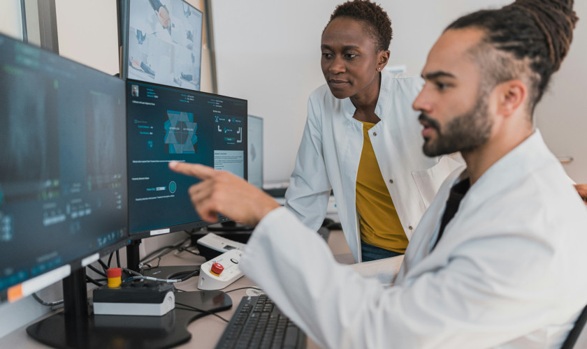
CT scanning is an indispensable imaging technique in modern medicine as it allows clinicians to have a fast, clear view on the patient’s anatomy. Although the scans provide valuable information, the associated radiation dose risk marks the importance of carefully selecting optimal scan settings in order to generate the best images at lowest necessary dose.
In our group, we developed new automatic methods to evaluate different aspects of clinical image quality, allowing to quantify what’s visually observed in CT images. In this project, you will use this software (originally developed for chest CT) on a large cohort of patient images to check whether quality is ensured in current CT protocols, expand the use to other anatomies (e.g., head), identify outliers and investigate where gains in image quality can be achieved.
The student will learn about the physics behind medical imaging, evaluate quality on patient images and apply statistics to large amounts of data.
- The project runs from 12 January 2026 - 1 July 2026.
- Number of placements available: 1 per semester.
Prerequisits
- High-level undergraduate student.
- Minimum GPA 3.4
Faculty Department
The group is a part of the department ‘imaging and pathology’ of the faculty of medicine, and focuses on medical physics aspects. Most of our research projects start from clinical remarks and/or complaints of the radiologists in the university hospital.
Topics of research include: quality evaluation of chest CT for lung cancer screening, optimisation of breast cancer screening techniques, patient dosimetry and automated dose and quality monitoring.
PhD students and master thesis students work under the guidance of Prof. Hilde Bosmans and Prof. Nicholas Marshall. Opportunities include the close cooperation with the radiology department, access to emerging x-ray equipment and patient images, a powerful software environment for online patient dose monitoring, and a framework of automatic image quality evaluation on CT scans.

